
Fetal Medicine
FIRST TRIMESTER EXTENDED SCREENING
By the 12th – 13th week of pregnancy, almost all organs of the fetus have developed, so that about two thirds of general developmental disorders that might develop can be detected with the help of modern, high resolution ultrasound devices. The first trimester extended screening at our clinic enables the detection of 95-97% of the most common severe chromosomal disorders (Down, Edwards, or Patau syndrome). However, only part of the screening focuses on chromosomal disorders. At this point, several other anatomical disorders can be detected, as well. During the first trimester extended screening, particular attention is paid to the screening of an embryo’s potential heart and cardiovascular disorders, as at this point – with the help of appropriate expertise – 90% of these severe disorders can already be detected. If necessary, we also provide genetic consultation.
The first trimester extended screening allows for the detection of more frequently occurring disorders, affecting the following organs and organ systems:
– missing skull bone
– upper jaw bone disorder (orofacial cleft), eye disorders (cyclopia)
Central nervous system and spine
– disorders of the brain hemispheres‘developments
– neural tube defects, spina bifida
Limbs
– anomaly in the number or form of arms and feet (e.g. missing limbs, clubfoot)
Chest and abdominal organs, abdominal wall
– anomaly in the form of the chest, open abdominal wall, umbilical hernia, diaphragmatic hernia
Kidneys, bladder
– renal pelvis dilatation, anomalies of the bladder
Heart and major vessels (approximately 90% of anomalies can be detected)
– formal, situational, and size abnormalities of the heart
– disorders of the heart chambers and atria, missing septa dividing the chambers
– disorders of major blood vessels (stenosis of the aorta or of the pulmonary artery, transposition of the great arteries, etc.)
– functional disorders of atrio-ventricular valves
– ultrasound signs of circulatory failure (pericardial fluid collection, chest and abdominal fluid)
Placenta
– disorder of the connection of the placenta to the uterine wall (low-lying placenta, hematoma)
Uterine wall and ovaries
– myoma, ovarian cysts
Twin pregnancies
– determination of chorionicity, screening of monochonionic twin pregnancies
– management of twin-to-twin transfusion syndrome (TTTS)
Ultrasound screening for abnormalities which increase the risk of chromosomal disorders and heart development defects
– nuchal translucency (NT)
– examination of missing fetal nasal bone (NB)
– tricuspid insufficiency (TR)
– abnormal ductus venosus flow (DV)
SECOND TRIMESTER ANATOMICAL SCREENING
Thanks to the growth of the fetus, by the second trimester (19th – 20th week), the ultrasound examination allows for the detection of 90% of developmental disorders and nearly 100% of severe heart defects. During the exam, in addition to the examination of the parameters indicating the fetus’ size, we do a 3D exam of the brain and facial bones, and the central nervous system can be examined in detail for the first time. Now spine, limbs – including fingers, chest, lungs, heart and major blood vessels, health of the abdominal wall, and the organs of the abdomen (stomach, bowels, gall bladder, liver, kidneys, bladder), navel and umbilical cord can be thoroughly examined and shown on 40 planes, which we look at during all exams. In addition, we also search for signs of specific abnormalities enhancing the risk of Down syndrome.
Disorders that can be screened for, listed by organ systems (the following list is non-exhaustive):
– morphological variations of the skull
– disorders of the cerebral hemispheres, the cerebellum and the water spaces around the brain / e.g. Dandy-Walker malformation, ventriculomegaly, hydrocephalus/
– shape and positional deviations of the eye
– abnormalities of the lips, hard palate, differences in the facial profile (wolf throat, rabbit cheeks, nasal bone underdevelopment)
– spinal column and disorders of the formation of the vertebrae (open spine, ossification disorders)
– anomaly in the number or form of arms and feet (e.g. missing limbs, clubfoot)
– abnormalities of the position, shape and size of the heart
– heart rhythm disorders
– deviations of ventricles and atria, lack of septa between heart cavities
– abnormalities of the great arteries (aorta, pulmonary artery, pulmonary veins), e.g. transposition of the great vessels, tetralogy of Fallot, aortic arch abnormalities, etc.)
– functional disorders of atrio-ventricular valves
– ultrasound signs of circulatory failure (pericardial fluid collection, chest and abdominal fluid)
– umbilical hernia
– small intestinal narrowing/obstruction, intestinal dilatation
– calcification of the liver
– underdevelopment/absence of kidneys, cystic degeneration of kidneys
– enlargement of the renal pelvis and urethra, abnormalities of the urinary bladder
– disorder of the connection of the placenta to the uterine wall (low-lying placenta, hematoma)
– e.g. liver, kidney, adrenal gland, teratomas, etc.
3RD TRIMESTER STATUS SCREENING
The third trimester ultrasound examination (28th – 31st week) focuses on the fetus’ growth and development inside the uterus. We will check umbilical and cerebral circulation, in order to check the functioning of the placenta. Considering that the majority of developmental disorders can already be detected during the second trimester ultrasound examination, anomalies detected at this point are generally of lesser significance. However, it can be important to know about them, because they might need to be treated and monitored after birth (smaller heart defects, renal anomalies, etc.)
GENETIC ADVICE
Already during the first semester, mothers-to-be receive genetic consultation if it is advised. During this consultation, we inform about the prevalence of chromosomal abnormalities and about the different genetic examinations that can be done.
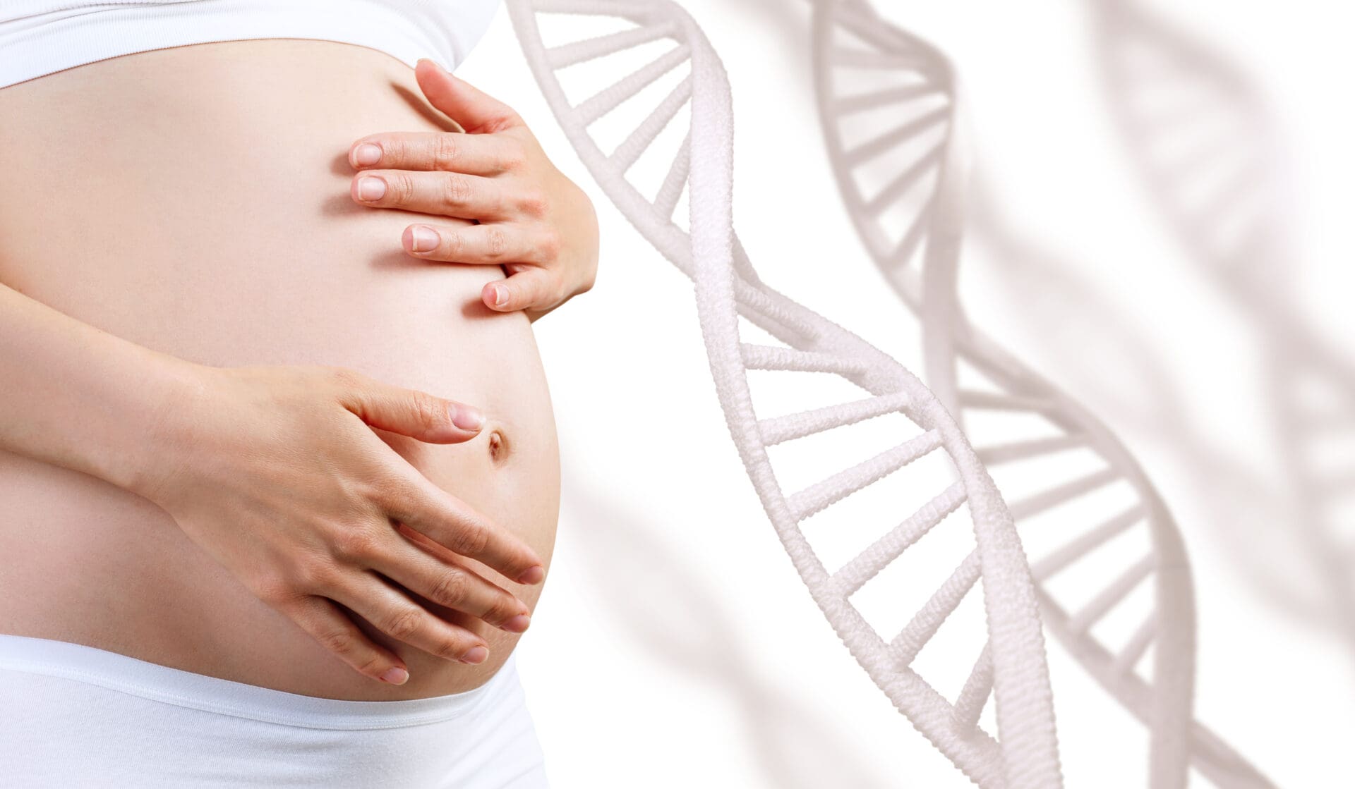
Our clinic offers the most up-to-date and modern genetic tests to everybody: the so-called non-invasive prenatal testing, or NIPT which enables to screen certain severe chromosomal disorders (Down, Edwards or Patau syndrome, sex chromosome disorders, and the deletion of smaller parts of certain chromosomes called microdeletion) with an accuracy of 99.5%, without endangering the embryo/fetus. Hence this testing does not bear any risk of miscarriage. The testing is based on the examination of the cell-free DNA of the embryo/fetus circulating in the mother’s blood (more precisely, DNA from the placenta cells). Non-invasive prenatal testing can be done at any time between the 9th and the 20th week with the same efficacy. It is advised, though, to choose the earliest date possible. The test results can be expected to be ready within 8-12 working days.